You’re staring at a microscopic blob. Is it a plant? Is it an animal? Honestly, if you’re looking at a poorly drawn diagram in a 1990s textbook, it’s probably just a mess of lines and circles. But getting plant and animal cell labelling right isn't just about passing a biology quiz. It’s about understanding the fundamental machinery of life. Look, cells are messy. They aren’t the perfect squares or circles you see in infographics. They are crowded, salty, vibrating factories.
Most people think the only difference is that plants have "green bits" and animals don't. That’s a massive oversimplification. If you really want to nail the identification, you have to look at the structural integrity and the storage systems.
The Rigid Reality of the Plant Cell
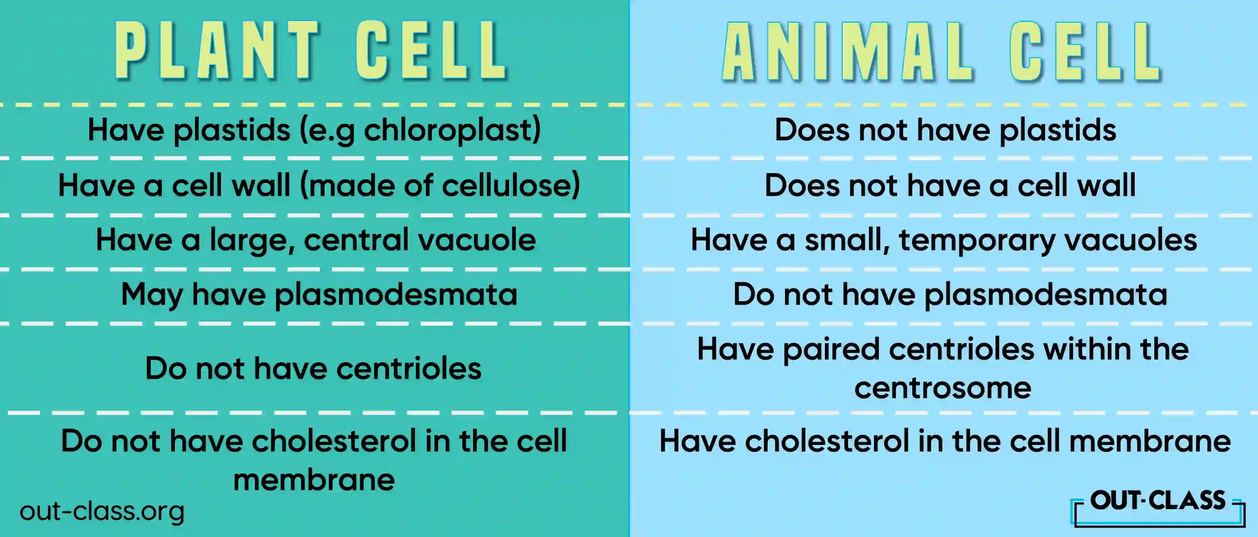
Plants are the architects of the cellular world. They can’t run away from a predator or move into the shade when it gets too hot, so they’ve evolved to be sturdy. When you start plant and animal cell labelling, the first thing your eye should hit is the cell wall. It’s thick. It’s made of cellulose. It’s why celery sticks crunch when you bite them.
Inside that wall, you’ve got the plasma membrane, which is basically the gatekeeper. But the real "wow" factor in a plant cell is the large central vacuole. Imagine a giant water balloon taking up 90% of the room. That’s the vacuole. It provides turgor pressure. Without it, the plant wilts and looks sad.
- Chloroplasts: These are the solar panels. They contain chlorophyll. They’re usually oval-shaped with stacks inside called thylakoids.
- Cell Wall: The outermost layer. It’s tough. It’s structural.
- Plasmodesmata: These are tiny channels. They let cells talk to each other through the walls. Most basic diagrams forget these entirely, but they are vital for nutrient transport.
Why Animal Cells Are The Shape-Shifters
Animal cells are the nomads. Since we have skeletons and muscles to hold us up, our individual cells don't need a heavy brick wall around them. This makes them squishy and irregular. When you’re doing plant and animal cell labelling for an animal cell, you’ll notice it looks way more chaotic.
There is no cell wall here. You just have the plasma membrane. It’s fluid. Think of it like a mosaic that’s constantly moving. One thing that usually trips students up is the centrosome. These contain centrioles—little pasta-shaped structures—that help with cell division. Plants mostly don't have these, or at least not in the same way.
Then there are the lysosomes. These are the "suicide bags" or garbage disposals of the cell. They’re packed with enzymes to break down waste. While plants have vacuole-like structures that do similar work, the lysosome is the classic hallmark of the animal cell diagram.
The Stuff They Both Have (The Universal Parts)
Don’t get it twisted—they have a lot in common. Both are eukaryotic. This means they both keep their DNA in a high-security vault called the nucleus.
The nucleus is the brain. It’s got the nucleolus inside, which is where ribosomes are born. Then you have the Endoplasmic Reticulum (ER). The "Rough" ER is covered in ribosomes and looks like sandpaper. It’s a protein factory. The "Smooth" ER is more about lipids and detoxing.
And we can't forget the mitochondria. The powerhouse? Yeah, everyone knows the meme. But specifically, it’s where ATP—the cellular currency of energy—is minted through oxidative phosphorylation. Both plants and animals need energy, so both have mitochondria. A common mistake in plant and animal cell labelling is thinking plants only have chloroplasts. Nope. They have both. They make the food, then they "burn" it for energy just like us.
The Cytoplasm vs. Cytosol Debate
People use these terms interchangeably. They shouldn’t. The cytosol is the liquid—the jelly. The cytoplasm is the entire area inside the membrane including the organelles but excluding the nucleus. It’s a subtle distinction, but if you’re aiming for expert-level accuracy, it matters.
Pro-Tips for Accurate Labelling
If you're actually sitting down to label a diagram, follow these "unspoken" rules of biological illustration:
- Don't cross your lines. It makes the diagram unreadable. Use a ruler.
- Keep labels to one side. Usually, the right side is standard.
- Use the correct terminology. It’s not a "green thingy," it’s a chloroplast.
- Look at the scale. If the vacuole is small, you’re likely looking at an animal cell. If it’s huge, it’s a plant.
Misconceptions That Will Sink Your Grade
One big myth is that animal cells are always round. They aren't. Think about a neuron (nerve cell). It’s long, spindly, and looks like a tree. Or a muscle cell, which is long and fibrous. The "round" animal cell is just a convenient lie we tell middle schoolers to make things easier.
Another one? The idea that plants don't have a cytoskeleton. They absolutely do. They have microtubules and actin filaments that help move things around inside the cell. Just because they have a wall doesn't mean they're static inside.
Real-World Application: Why This Matters
Why do we care about plant and animal cell labelling? Well, medicine and agriculture depend on it. When scientists develop herbicides, they target things only plant cells have—like the enzymes involved in making the cell wall or specific parts of the chloroplast. This way, the chemicals kill the weeds but don't hurt the humans (animal cells) applying them.
In medicine, understanding the animal cell membrane is how we design drugs. If a drug needs to get into a cell, it has to be able to bypass the phospholipid bilayer. Knowing which receptors are on the surface—the "labels" of the cell—is the difference between a life-saving treatment and a dud.
Actionable Next Steps
To truly master this, don't just look at a finished diagram. Build one.
- Draw it from memory: Start with the nucleus and work your way out. If you get stuck, that’s your "knowledge gap."
- Use the "Function First" Method: Instead of memorizing names, memorize what the cell needs to do (Eat, Breathe, Waste, Structure) and then find the organelle that does it.
- Compare under a microscope: If you have access to one, look at onion skin (plant) versus a cheek swab (animal). You'll see the "bricks" of the onion vs. the "fried eggs" of the cheek cells immediately.
- Color code your notes: Use green for plant-exclusive structures and red or blue for the shared ones. It creates a visual anchor in your brain.
The key to plant and animal cell labelling is recognizing that these aren't just static images. They are blueprints for how life operates. Once you see the logic behind the structure—why the wall exists or why the mitochondria are everywhere—the labels start to make sense on their own.